PTEN FISH Probe
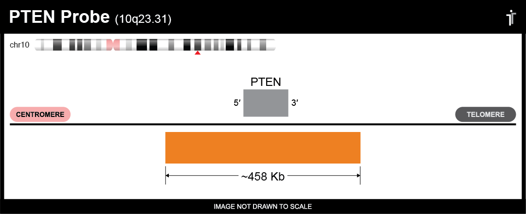
Our PTEN probe is designed to detect PTEN amplifications and deletions. The probe comes labeled in orange, but can be customized to meet your needs.

** This product is for in vitro and research use only. This product is not intended for diagnostic use.

| SKU | Test Kits | Buffer | Dye Color | Order Now |
|---|---|---|---|---|
| PTEN-20-OR (Standard Design) | 20 (40 μL) | 200 μL |

|
|
| PTEN-20-RE | 20 (40 μL) | 200 μL |

|
|
| PTEN-20-AQ | 20 (40 μL) | 200 μL |

|
|
| PTEN-20-GR | 20 (40 μL) | 200 μL |

|
|
| PTEN-20-GO | 20 (40 μL) | 200 μL |

|
Gene Summary
This gene was identified as a tumor suppressor that is mutated in a large number of cancers at high frequency. The protein encoded by this gene is a phosphatidylinositol-3,4,5-trisphosphate 3-phosphatase. It contains a tensin like domain as well as a catalytic domain similar to that of the dual specificity protein tyrosine phosphatases. Unlike most of the protein tyrosine phosphatases, this protein preferentially dephosphorylates phosphoinositide substrates. It negatively regulates intracellular levels of phosphatidylinositol-3,4,5-trisphosphate in cells and functions as a tumor suppressor by negatively regulating AKT/PKB signaling pathway. The use of a non-canonical (CUG) upstream initiation site produces a longer isoform that initiates translation with a leucine, and is thought to be preferentially associated with the mitochondrial inner membrane. This longer isoform may help regulate energy metabolism in the mitochondria. A pseudogene of this gene is found on chromosome 9. Alternative splicing and the use of multiple translation start codons results in multiple transcript variants encoding different isoforms. [provided by RefSeq, Feb 2015]
Gene Details
Gene Symbol: PTEN
Gene Name: Phosphatase And Tensin Homolog
Chromosome: CHR10: 89623194-89728532
Locus: 10q23.31
FISH Probe Protocols
| Protocol, Procedure, or Form Name | Last Modified | Download |
|---|